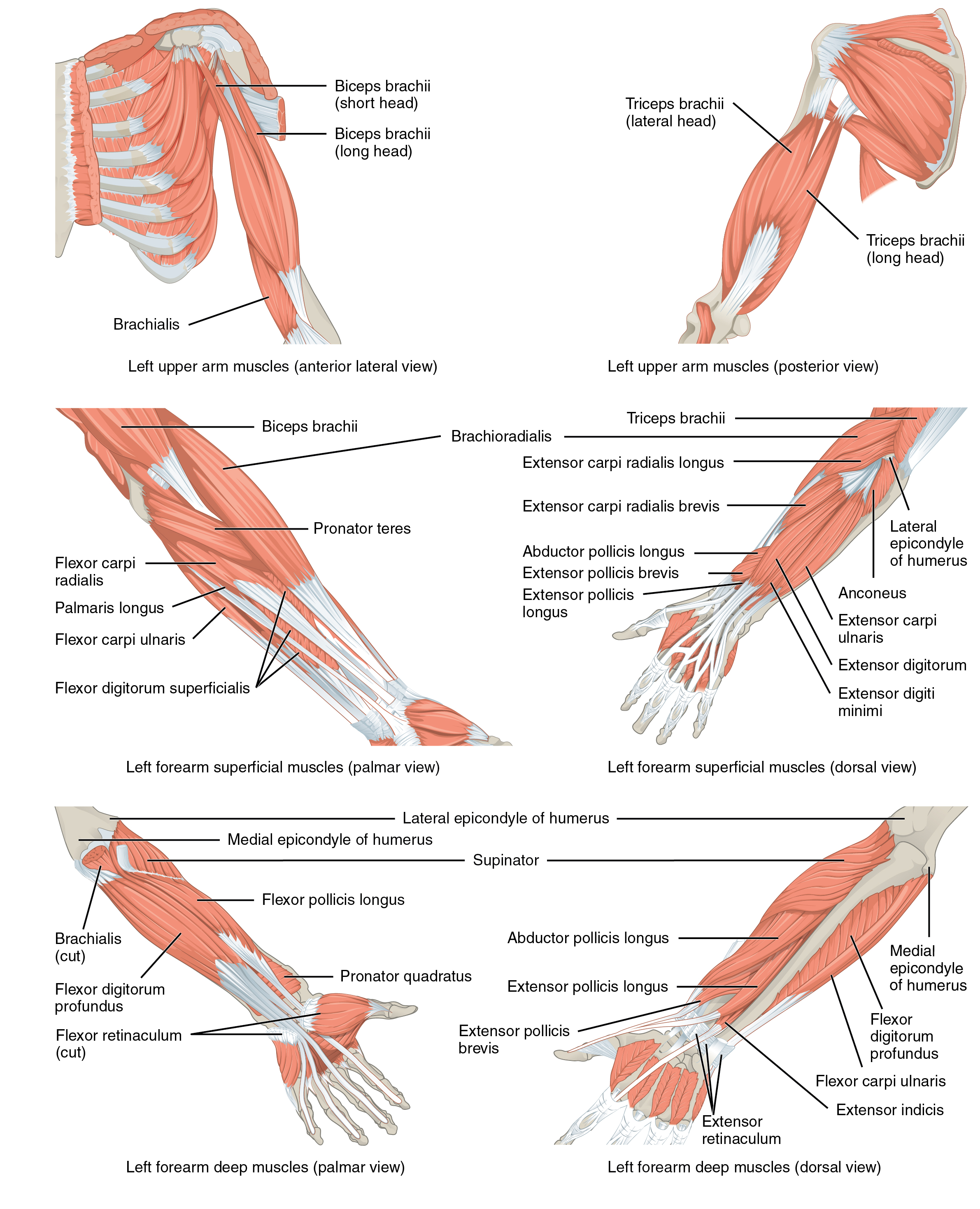
Diagram Of The Muscles In The Forearm : How to use Resisted and Assisted Sprint Training in Swimming / Some of the muscles also function to supinate the forearm, a rotatory movement at the elbow wrist axis which brings the palms towards the sky.
Diagram Of The Muscles In The Forearm : How to use Resisted and Assisted Sprint Training in Swimming / Some of the muscles also function to supinate the forearm, a rotatory movement at the elbow wrist axis which brings the palms towards the sky.. The muscles of the upper arm are responsible for the flexion and extension of the forearm at the elbow joint. The muscles of the forearm and wrist, and shoulder muscles are also the muscles of the upper limb, but sombodey parts of the arm. Longus, brevis, longus, brevis (longus is lateral to brevis). The anterior forearm muscles are divided into 3 muscular layers ; The forearm is a mass of some 20 different muscles.
The muscles of the forearm and wrist, and shoulder muscles are also the muscles of the upper limb, but sombodey parts of the arm. Remembering the action of each one can be quite difficult. In the anterior compartment, they are split into three categories: Try labeling diagrams and worksheets as additional learning aids. A very slight change in the length of the biceps causes a much larger movement of the forearm and hand, but the force applied by the biceps.
Muscles of the Pectoral Girdle and Upper Limbs · Anatomy ... from philschatz.com In the anterior compartment, they are split into three categories: Inflammation of this region caused by repetitive. 2, ulna, 3, biceps muscle; Strength training exercises are common ways to increase the size and overall strength of the major muscles in the arms. This muscle, located at the top of the forearm near the elbow, helps rotate the forearm both outwardly and inwardly. It is a functionally important muscle that contains two heads. Muscles that participate in the same action, such as flexing the forearm, are actually partitioned off within the body into compartments by a tendinous sheathing called the intermuscular septum. Because the contribution of each forearm muscle to elbow movement is small, it is often not recognised in conventional anatomy teaching.
Superficial muscles of the posterior forearm:
I made an entire tutorial dedicated to drawing the forearms with anatomical detail, it can be fond here. Diagram the movements of the humerus muscles that act on the forearm. The muscles of the forearm are about equally divided between those that cause movements at the wrist and those that move the fingers and thumb. Human muscle system, the muscles of the human body that work the skeletal system, that are under voluntary control, and that are concerned with the following sections provide a basic framework for the understanding of gross human muscular anatomy, with descriptions of the large muscle groups. Learn vocabulary, terms and more with flashcards, games and other study tools. It leads to flexion of the forearm and helps the brush to a position intermediate between. Try labeling diagrams and worksheets as additional learning aids. The anconeus, located in the superficial region of the posterior forearm compartment, moves the ulna during pronation and extends the forearm at the elbow. It is a functionally important muscle that contains two heads. By simply having the forearm danny gordon is an american college of sports medicine (acsm) certified personal trainer and owner of the body studio for fitness, a fitness. The flexor pollicis longus is situated on the radial side of the forearm, lying in the same plane as the preceding. Superficial muscles of the posterior forearm: This muscle, located at the top of the forearm near the elbow, helps rotate the forearm both outwardly and inwardly.
This layer contains only one muscle, the flexor digitorum. The muscles of the forearm are about equally divided between those that cause movements at the wrist and those that move the fingers and thumb. Longus, brevis, longus, brevis (longus is lateral to brevis). Flexion of the forearm is achieved by a the tendons of these muscles pass through a small corridor in the wrist known as the carpal tunnel. I've just switched over to a diagram to show you this muscle.
Shoulder Muscles Anatomy, Diagram & Function | Body Maps from post.healthline.com It is a functionally important muscle that contains two heads. Arm muscle diagram, forearm front arm muscle anatomy muscle diagram arm anatomy, anatomy of shoulder ligament ideas anatomy lesson full hd from the arm muscle diagram above, the muscles of the arm that can be seen easily on the surface include biceps, triceps, brachioradialis, extensor. The superficial extensors of the forearm are the brachioradialis, extensor carpi radialis longus, anconeus, extensor carpi radialis brevis, extensor carpi ulnaris, extensor digitorum and extensor digiti minimi. Strength training exercises are common ways to increase the size and overall strength of the major muscles in the arms. Learn vocabulary, terms and more with flashcards, games and other study tools. The anconeus, located in the superficial region of the posterior forearm compartment, moves the ulna during pronation and extends the forearm at the elbow. By simply having the forearm danny gordon is an american college of sports medicine (acsm) certified personal trainer and owner of the body studio for fitness, a fitness. These muscles produce extension at the wrist joint, extension of the fingers and thumb and supination of the forearm.
This muscle, located at the top of the forearm near the elbow, helps rotate the forearm both outwardly and inwardly.
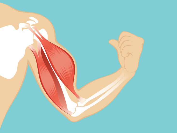
A very slight change in the length of the biceps causes a much larger movement of the forearm and hand, but the force applied by the biceps. This is the most medial of the superficial flexor muscles in the forearm. Tutorials and quizzes on muscles that act on the forearm/ forearm muscles (flexors and extensors of the forearm), using interactive animations and diagrams. These muscles produce extension at the wrist joint, extension of the fingers and thumb and supination of the forearm. Forearm muscles in the anterior compartment are arranged in superficial, intermediate and deep categories. The muscles of the upper arm are responsible for the flexion and extension of the forearm at the elbow joint. Longus, brevis, longus, brevis (longus is lateral to brevis). Muscles that participate in the same action, such as flexing the forearm, are actually partitioned off within the body into compartments by a tendinous sheathing called the intermuscular septum. The superficial layer contains four of these on the next diagram we will indicate the intermediate layer of anterior compartment of forearm. Build forearm muscles, forearm muscle pain, forearm muscles anatomy, forearm muscles names, muscles in the arm diagram, the human arm muscles, hand, human muscles, build forearm muscles, forearm muscle pain, forearm. Remembering the action of each one can be quite difficult. Inflammation of this region caused by repetitive. In the anterior compartment, they are split into three categories:
A deep layer , intermediate layer and superficial layer. Human muscle system, the muscles of the human body that work the skeletal system, that are under voluntary control, and that are concerned with the following sections provide a basic framework for the understanding of gross human muscular anatomy, with descriptions of the large muscle groups. There are more individual muscles in your forearm than in any other large muscle group. The antibrachial or forearm muscles may be divided into a volar and a dorsal group. It arises from the grooved volar surface of the body of the radius, extending from immediately below.
Arm Muscle Diagrams from www.101diagrams.com The term forearm is used in anatomy to distinguish it from the arm. The muscles of the upper arm are responsible for the flexion and extension of the forearm at the elbow joint. Learn vocabulary, terms and more with flashcards, games and other study tools. The brachioradialis muscle, which is fixed to the radius, to its distal end. The anterior forearm muscles are divided into 3 muscular layers ; There are many muscles in the forearm. Try labeling diagrams and worksheets as additional learning aids. The superficial extensors of the forearm are the brachioradialis, extensor carpi radialis longus, anconeus, extensor carpi radialis brevis, extensor carpi ulnaris, extensor digitorum and extensor digiti minimi.
In the anterior compartment, they are split into three categories:
The superficial extensors of the forearm are the brachioradialis, extensor carpi radialis longus, anconeus, extensor carpi radialis brevis, extensor carpi ulnaris, extensor digitorum and extensor digiti minimi. Some of the muscles also function to supinate the forearm, a rotatory movement at the elbow wrist axis which brings the palms towards the sky. Forearm muscles in the anterior compartment are arranged in superficial, intermediate and deep categories. The forearm is a mass of some 20 different muscles. Strength training exercises are common ways to increase the size and overall strength of the major muscles in the arms. Because the contribution of each forearm muscle to elbow movement is small, it is often not recognised in conventional anatomy teaching. 11 photos of the forearm muscles diagram structure. I've just switched over to a diagram to show you this muscle. 4, attachment… the muscles of the back forearm. Superficial muscles of the posterior forearm: Tutorials and quizzes on muscles that act on the forearm/ forearm muscles (flexors and extensors of the forearm), using interactive animations and diagrams. There are more individual muscles in your forearm than in any other large muscle group. The forearm is the region of the upper limb between the elbow and the wrist.


0 Comments